
Paraffin Tissue Block
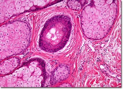
What is Tissue?
In biology, tissue is a cellular organizational level between cells and a complete organ. A tissue is an ensemble of similar cells and their extracellular matrix from the same origin that together carry out a specific function. Organs are then formed by the functional grouping together of multiple tissues. The study of tissue is known as histology or, in connection with disease, histopathology. The classical tools for studying tissues are the paraffin block in which tissue is embedded and then sectioned, the histological stain, and the optical microscope. In the last couple of decades, developments in electron microscopy, immunofluorescence, and the use of frozen tissue sections have enhanced the detail that can be observed in tissues. With these tools, the classical appearances of tissues can be examined in health and disease, enabling considerable refinement of medical diagnosis and prognosis.
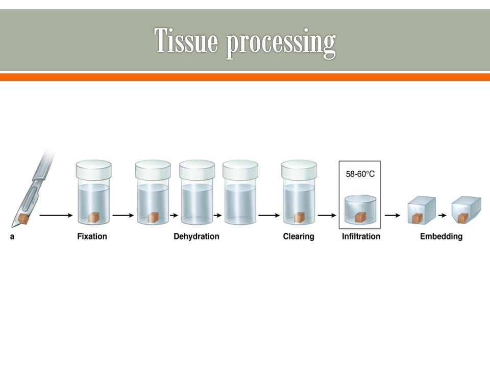
Fixation
Fixation is performed to preserve the structure of the tissue. This process provides rigidity to the tissue, making it easier to section. There are a variety of forms of fixation, including “fresh frozen,” where the tissue is not actually placed in fixative. Common fixatives used include formaldehyde (formalin) and glutaraldehyde. Ethanol, acetone, and paraformaldehyde are often used for cryopreserved specimens. While samples are often fixed before cryopreservation, this is not always necessary.
The choice of fixative depends on the staining technique (histological staining, immunohistochemistry, in situ hybridization) to be used later. Most histological staining methods, but not all, allow the use of formalin. Immunohistochemistry can be performed on paraffin/plastic embedded or cryopreserved specimens that have been fixed, but some antibodies will not be able to bind to the target molecules after the formalin fixation and paraffin embedding processes.
Once placed in the fixative, covalent bonds are formed within the amine groups of the tissue, crosslinking it. A tissue sample is fixed for only a short period of time, usually overnight, as longer fixation times may cause artifacts to be apparent at high levels of magnification. Once samples have been fixed, they can be kept indefinitely at room temperature. Once fixation has been completed, the sample is embedded prior to sectioning.
Embedding
Before sectioning, tissue samples are embedded in a material with similar mechanical properties. This step allows the tissue to be cut easily.
If a tissue is to be sectioned with a microtome, it can be embedded in paraffin, or in plastic resin if the tissue is particularly hard. After fixation, tissues that are to be paraffin-embedded are dehydrated by first using graded ethanol solutions, then graded xylene solutions, then finally liquid paraffin. The graded solutions gradually expose the sample to changes in hydrophobicity, minimizing damage to cells. After a short time in the liquid paraffin, the tissue is placed into a mold with more paraffin. The wax is allowed to solidify, forming a block that can be held in a microtome.
Paraffin infiltration
In this procedure, tissue is dehydrated through a series of graded ethanol baths to displace the water, and then infiltrated with wax. The infiltrated tissues are then embedded into wax blocks. Once the tissue is embedded, it is stable for many years.
The most commonly used waxes for infiltration are the commercial paraffin waxes. A paraffin wax is usually a mixture of straight chain or n-alkanes with a carbon chain length of between 20 and 40; the wax is a solid at room temperature but melts at temperatures up to about 65°C or 70°C. Paraffin wax can be purchased with melting points at different temperatures, the most common for histological use being about 56°C–58°C, At its melting point it tends to be slightly viscous, but this decreases as the temperature is increased. The traditional advice with paraffin wax is to use this about 2°C above its melting point. To decrease viscosity and improve infiltration of the tissue, technologists often increase the temperature to above 60°C or 65°C in practice to decrease viscosity.
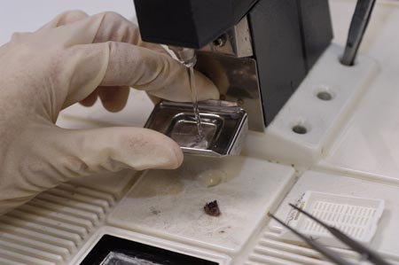
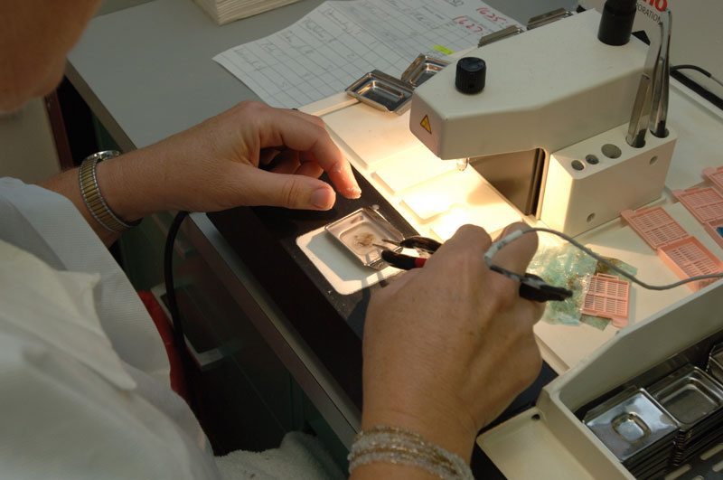
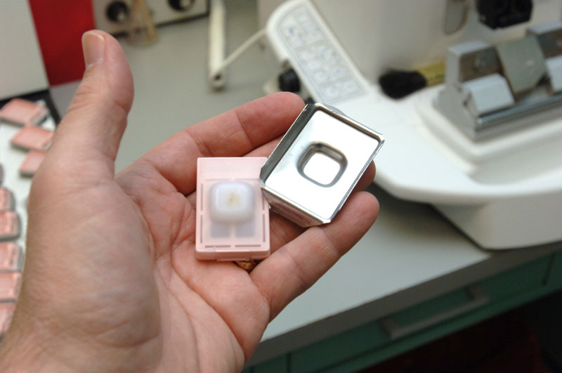
Embedding tissues in paraffin blocks (Paraffin Tissue Block)
Tissues processed into paraffin will have wax in the cassettes; in order to create smooth wax blocks, the wax first needs to be melted away placing the entire cassette in 58°C paraffin bath for 15 minutes. Turn the heat block on to melt the paraffin one hour before adding the tissue cassettes.
- Open cassette to view tissue sample and choose a mold that best corresponds to the size of the tissue. A margin of at least 2 mm of paraffin surrounding all sides of the tissue gives best cutting support. Discard cassette lid.
- Put small amount of molten paraffin in mold, dispensing from paraffin reservoir.
- Using warm forceps, transfer tissue into mold, placing cut side down, as it was placed in the cassette.
- Transfer mold to cold plate, and gently press tissue flat. Paraffin will solidify in a thin layer which holds the tissue in position.
- When the tissue is in the desired orientation add the labeled tissue cassette on top of the mold as a backing. Press firmly.
- Hot paraffin is added to the mold from the paraffin dispenser. Be sure there is enough paraffin to cover the face of the plastic cassette.
- If necessary, fill cassette with paraffin while cooling, keeping the mold full until solid.
- Paraffin should solidify in 30 minutes. When the wax is completely cooled and hardened (30 minutes) the paraffin block can be easily popped out of the mold; the wax blocks should not stick. If the wax cracks or the tissues are not aligned well, simply melt them again and start over.
The tissue and paraffin attached to the cassette has formed a block, which is ready for sectioning.Tissue blocks can be stored at room temperature for years.
Sectioning
Samples embedded in paraffin are first mounted in a microtome. The microtome holds a sharp blade and is controlled by a crank that is turned to bring the paraffin block closer to the blade. As the crank is turned further, the blade cuts slices of paraffin, which containing tissue. The microtome can be adjusted for width and angle of cut. After sectioning, the slices can be placed on a slide.